Advantages
- Unprecedented coverage: 12mm FOV covers entire corneal surface—2× larger than any quantitative interferometer
- Sub-micron precision: ~0.38 μm resolution enables detailed tear film layer analysis
- Multimodal platform: Integrates tear film interferometry with OCT in single device
- Spatial heterogeneity mapping: Identifies localized thin zones missed by point measurements
Summary
Dry eye disease affects over 344 million people globally, yet clinicians lack tools for precise, quantitative diagnosis. Current devices either provide quantitative measurement of limited corneal regions OR broad coverage with only qualitative assessment. No commercial device maps tear film thickness quantitatively across the entire corneal surface—critical for detecting localized dysfunction that triggers symptoms.
Our researchers developed a multimodal imaging platform that achieves sub-micron resolution (~0.38 μm) across a 12mm diameter field of view—covering the entire exposed cornea. A novel curved focal plane objective lens and proprietary algorithm enable quantitative tear film dynamics mapping that was previously impossible, providing objective data for diagnosis and treatment monitoring.
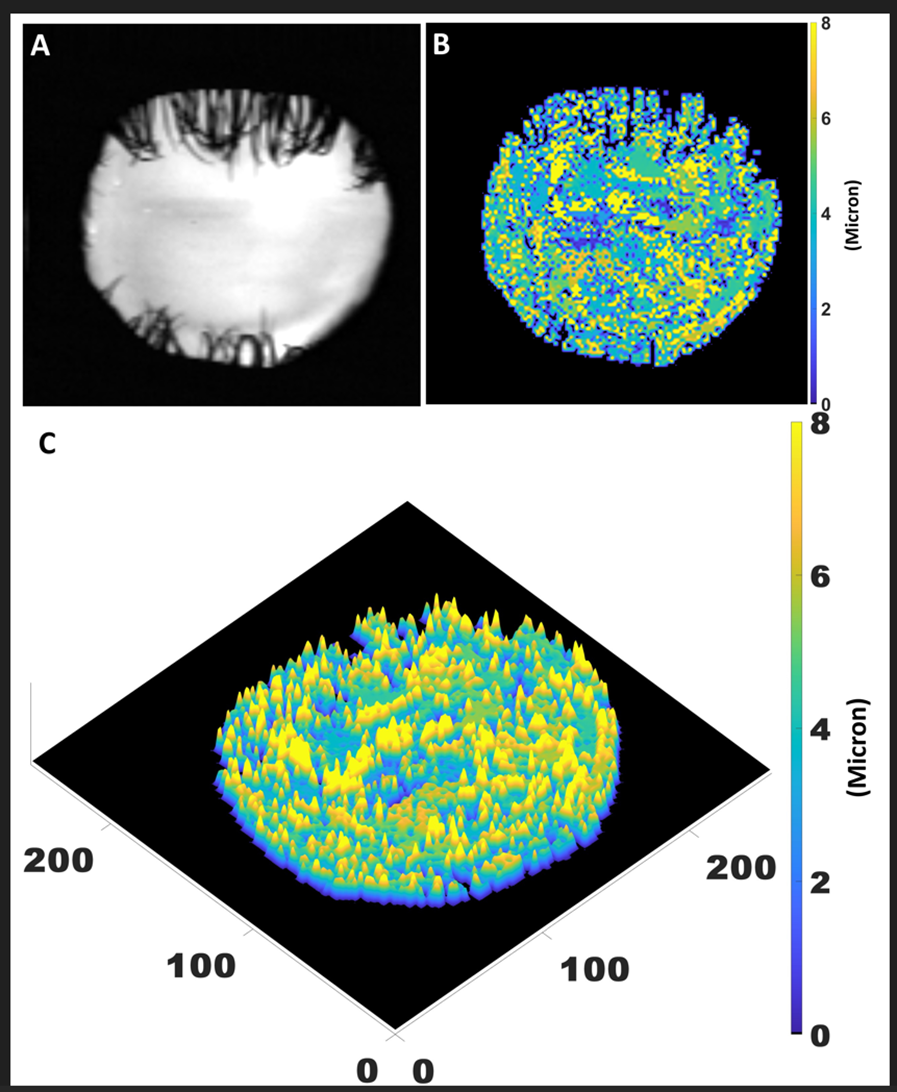
Fig. 1. The corneal surface of one human subject. (A) The image based on the reflected intensity; (B) the thickness map of precorneal tear film (PCTF) based on the color bar, and (C) the surface plot based on its thickness value.
Desired Partnerships
- License
- Sponsored Research
- Co-Development