Advantages:
- This study discovered a potential novel mechanism of cardiac dysfunction in Friedreich's ataxia (FA) by uncovering dysregulation in key calcium-handling proteins and mitochondrial dysfunction
- The study correlates data from an FA mouse model (FXN-KO) with human clinical manifestations, linking frataxin deficiency to cardiac dysfunction in Friedreich's ataxia
- These findings pave the way for targeted therapies in Friedreich's ataxia
Summary:
Friedreich’s ataxia (FA) is an inherited neurological disorder caused by an expansion of the DNA triplet (GAA) in the first intron of the Frataxin gene (FXN), which decreases the expression of the frataxin protein, a mitochondrial protein. Although FA is considered a neurodegenerative disorder, over 59% of patients develop heart failure. The molecular mechanisms underlying the cardiac dysfunction in FA remains poorly understood.
Our researchers identified how the abnormal Ca2+ handling machinery and the associated mitochondrial dysfunction can cause left ventricular contractile failure and lead to perpetuated cardiac dysfunction in FA. Using a frataxin knock-out mouse model and human cardiac tissues from FA patients they found that the major calcium-handling proteins, SERCA2a and Ryr2, were downregulated. This resulted in altered contractility kinetics, depolarized mitochondrial membrane potential, elevated reactive oxygen species (ROS), and decreased oxygen consumption rate (OCR), as well as impaired sarcomere and whole cell shortening.
The results of this study elucidate a potential novel mechanism of left ventricular contractile dysfunction in FA that is associated with reduced expression of calcium handling proteins and mitochondrial dysfunction, which provides important information for potential therapeutic targets.
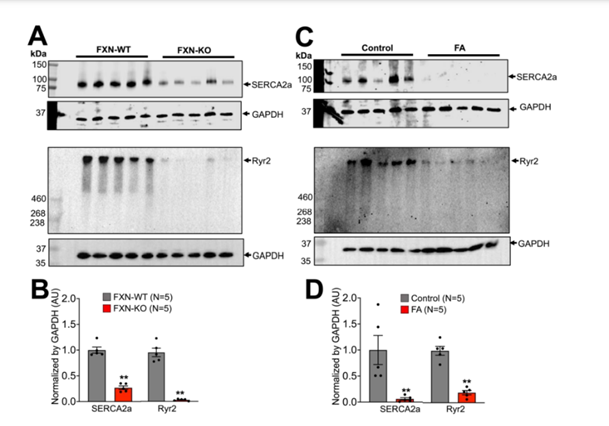
Western blot analysis of major Ca 2+ handling proteins levels in FA. Representative blots of sarco-endoplasmic reticulum ATPase 2a (SERCA2a), and ryanodine receptor (RyR2) in FXN-WT and KO (A), and control and FA (C) ventricular tissue samples. Barographs are quantification of SERCA2a and RyR2 normalized by GAPDH. SERCA2a and RyR2 were significantly decreased in FXN-KO compared to the WT (B) and in FA compared to the control (D). (** p < 0.01 vs. control or WT; unpaired 2-sample t-test). Bars are mean ± SEM.
Desired Partnerships:
• License
• Sponsored Research
• Co-Development