Advantages:
- Enhanced diagnostic accuracy for precise cancer cell identification.
- Complementary insights from both morphology and surface potential analysis.
- Label-free and minimally invasive, ensuring safety and sample integrity.
Background:
Breast and ovarian are the most frequent types of cancer among women worldwide. Morbidity and mortality of cancer are substantially decreased with early detection. Cytological
screening tests have decreased mortality. However, these methods have insufficient sensitivity and are time-consuming in both analysis and training of professionals with subjective manual diagnosis. More accurate tests may substantially decrease the cost and patient inconvenience.
Summary:
Recent advances in high-resolution biomedical imaging focusing on morphological, electrical, and biochemical properties of cells and tissues, scaling from cell clusters down to the molecular level, have improved cancer diagnosis. Multiscale imaging revealed high complexity that requires advanced data processing methods of multifractal analysis. USF researchers performed label-free multiscale imaging of surface potential variations in human ovarian and breast cancer cells using Kelvin probe force microscopy (KPFM). The advanced fractal and multifractal analysis techniques unveiled disparities in both cell morphology and surface potential. Notably, the comparative analysis between atomic force microscopy (AFM) and KPFM revealed a significant finding: KPFM consistently demonstrated superior efficacy in distinguishing between normal and cancer cells. The high spatial resolution allows for probing a broad range of scales, covering more than three orders of magnitude, from < 10 nm to tens of micrometers. The results reveal the potential of using multifractality as a new biomarker for cancer diagnosis. Furthermore, the surface potential imaging can be used in combination with morphological imaging for cancer diagnosis.
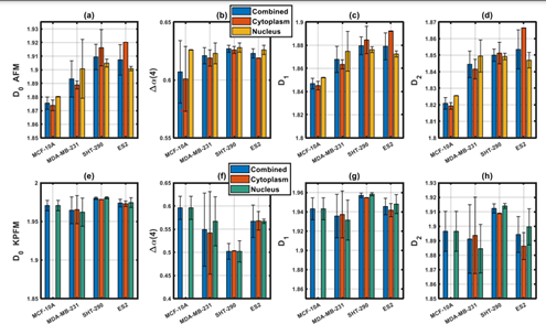
In Figure 1, we see different patterns between AFM and KPFM multifractal parameters. KPFM images of SHT290 cells show higher 𝐷0, 𝐷1, and 𝐷2 values compared to ES-2 cells, indicating a lower local fractal dimension in ovarian cancer cells versus normal endometrial cells. ES-2 cells exhibit greater variability in these parameters, suggesting increased heterogeneity. ES-2 cells also display higher multifractality in their surface potential, reflected in a notably higher average Δ𝛼(4) value (p < 0.05). There are no significant differences in the mean values of surface potential Δ𝛼(4) between MCF-10A and MDA-MB-231 cells, but MDA-MB-231 cells show more pronounced variations in Δ𝛼(4).
Desired Partnerships:
- License
- Sponsored Research
- Co-Development