Advantages:
• Detects and quantifies multiple discrete infarcts rather than a single total volume.
• Identifies very small (sub-milliliter) lesions often missed by standard solutions.
• Measures both absolute infarct volume and normalized burden (% of brain volume).
• Uses 3D connectivity and morphological analysis for more reliable lesion segmentation.
• Delivers rapid, automated analysis directly from standard MRI data.
Summary
Stroke diagnosis is highly time-sensitive, yet the complexity of 3D imaging data from CT and MRI can slow critical decision-making. Clinicians must interpret heterogeneous lesion patterns across multiple slices, a process that is time-consuming and often lacks quantitative precision. As a result, subtle variations in infarct size, distribution, and underlying etiology may be missed or incompletely characterized, limiting optimal treatment planning.
Our inventors developed an AI-guided imaging system for automated detection and 3D quantification of stroke lesions from diffusion‑weighted MRI (DWI). The platform uses a semi-automated MATLAB pipeline and high-performance computing to process DICOM data from MRI scans, segment infarct regions, and identify 3D-connected lesion volumes. Proof-of-concept studies demonstrate rapid detection of multiple infarcts—including sub‑milliliter lesions—and accurate measurement of both absolute and normalized infarct burden. These results support readiness for clinical integration, enabling real-time triage, quantitative assessment, and improved decision-making across acute care and longitudinal monitoring.
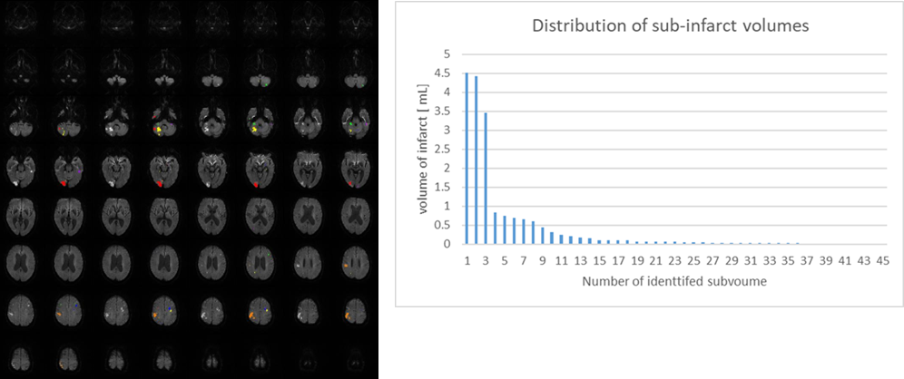
AI-Based 3D Quantification of Complex Multifocal Stroke:
AI-guided analysis of a complex cardioembolic stroke from diffusion-weighted MRI identifies 45 discrete infarct volumes and reconstructs them in 3D. The system automatically computes total infarct volume (18.8 mL) and relative infarct burden (1.46% of brain volume), while quantifying individual lesion sizes. This example demonstrates precise detection, multi-lesion segmentation, and scalable quantification of complex stroke patterns beyond conventional single-volume approaches.
Desired Partnerships
- License
- Sponsored Research
- Co-Development