Competitive Advantages
- Automatically provides measurements of pelvic region from MRI images
- More accurate than manual diagnosis
- Capable of differentiating between patients with & without POP
Summary
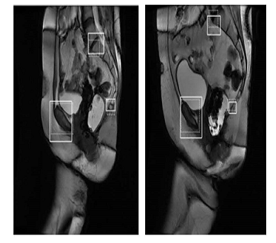
USF inventors have developed a novel system for the automated measurement and analysis of MRI images of the pelvic region. The new approach evaluates MRI images and presents clinical information related to the diagnosis of POP. The system can automatically identify important reference points in the pelvic region faster, more accurately, and with greater consistency compared with manual identification by experts. The system also provides a prediction model that is capable of telling the difference between patients with and without POP. This image analysis tool can be applied to the automated detection of features in MRI images for the diagnosis of POP as well as other diseases and internal damage where current clinical examination is inadequate.

Detection Accuracy: Solid Lines are Areas Detected by Presented Method, Dashed Lines are True Reference Areas
Desired Partnerships
- License
- Sponsored Research
- Co-Development